Pattern of sinonasal tumours presented in Shaheed Ziaur Rahman Medical College Hospital, Bogura, Bangladesh
Abstract
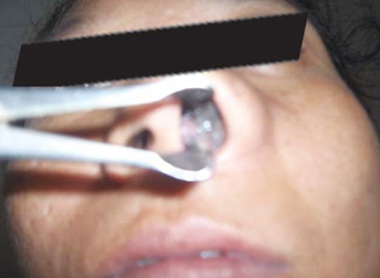
Introduction: Sinonasal tumors most commonly arise from the nasal cavity, followed by the maxillary and ethmoid sinus; sphenoid and frontal sinus tumors are both extremely rare entities. In the sinonasal complex, squamous cell carcinoma (SCC) is the most common histology, constituting 40 to 50% of all sinonasal malignancies. Objective: To assess the pattern of sinonasal tumors presented in Shaheed Ziaur Rahman Medical College Hospital, Bogura, Bangladesh. Methods: This is a retrospective study carried out in the Department of ENT & Head-Neck Surgery, Shaheed Ziaur Rahman Medical College Hospital, Bogura, Bangladesh from January to June 2022. It includes 52 cases of neoplastic sinonasal growths. All cases were thoroughly evaluated including history, head and neck examination including endoscopy, imaging and histopathological examinations. All the non-neoplastic cases were excluded from the study. Details of clinical presentation, examination, radiological and histopathological findings were recorded. Results: A total of 52 sinonasal tumors presented during the period. Out of which, 41 were benign and 12 were malignant tumors. Out of 41 benign tumors, inverted papilloma was the most common comprising 12 cases (22.6%) followed by squamous (epithelial) papilloma 11 cases (20.8%), hemangioma 10 cases (18.9%), osteoma and fibrous dysplasia each 2 cases (3.8%) and ossifying fibroma, pleomorphic adenoma and angiomyoma one case each (2%). Out of 12 malignant tumors, squamous cell carcinoma was the commonest malignancy observed in the study. They were four in number (7.5%), followed by basal cell carcinoma in three cases (5.7%) and malignant melanoma in two (3.8%). Adenocarcinoma, osteosarcoma and Rhabdomyosarcoma were each one in number (5.37%) (Table-1). Nasal blockage (94.3%), nasal discharge (66.04%), epistaxis (39.6%), hemifacial pain/pressure (34%) and facial fullness/external deformities, each (18.9%) were among the commonest presentation. Conclusion: The similarities of benign and malignant disorders at initial presentation may lead to a significant delay in the diagnosis of malignancy. Key indicators of malignancy such as cranial neuropathies and proptosis are uncommon at initial presentation and signify advanced disease. Neoplasms of the nasal cavity and paranasal sinuses are rare but require a high index of suspicion for diagnosis due to the overlapping presentation between benign and malignant ones.
Downloads
References
Haerle S.K., Gullane P.J., Witterick I.J., Zweifel C., Gentili F. Sinonasal carcinomas: Epidemiology, pathology, and management. Neurosurg. Clin. N. Am. 2013;24:39–49.
Turner J.H., Reh D.D. Incidence and survival in patients with sinonasal cancer: A historical analysis of population-based data. Head Neck. 2012;34:877–885.
Cooper J.S., Porter K., Mallin K., Hoffman H.T., Weber R.S., Ang K.K., Gay E.G., Langer C.J. National Cancer Database report on cancer of the head and neck: 10-year update. Head Neck. 2009;31:748–758. doi: 10.1002/hed.21022.
Razek A.A., Sieza S., Maha B. Assessment of nasal and paranasal sinus masses by diffusion-weighted MR imaging. J. Neuroradiol. 2009;36:206–211. doi: 10.1016/j.neurad.2009.06.001.
Sasaki M., Eida S., Sumi M., Nakamura T. Apparent diffusion coefficient mapping for sinonasal diseases: Differentiation of benign and malignant lesions. AJNR Am. J. Neuroradiol. 2011;32:1100–1106.
Dulguerov P, Jacobsen MS, Allal AS, Lehmann W, Calcaterra T. Nasal and paranasal sinus carcinoma: are we making progress? A series of 220 patients and a systematic review. Cancer 2001; 92: 3012-29.
Mills SE, Fechner RE. The nose, paranasal sinuses and nasopharynx. In: Sternberg SS, editor. Diagnostic Surgical Pathology. (3rd ed). Philadelphia: Lippincott Williams & Wilkins1999: 885-92.
Hernberg S, Westerholm P, Schultz-Larsen K, et al. Nasal and sinonasal cancer. Connection with occupational exposures in Denmark, Finland and Sweden. Scand J Work Environ Health 1983; 9: 315-26.
Osguthorpe JD. Sinus Neoplasia. Arch Otol Head Neck Surg 1994; 120: 19-25.
Hanna E, Westfall CT. Cancer of nasal cavity, paranasal sinuses and orbit. In Myers EN, et al, editor. Cancer of the head and neck. (4th ed). Philadelphia: Elsevier 2003: 163.
Golden berg D, Golz A, Fradis M, Mârtu D, Netzer A, Joachims HZ. Malignant tumours of the nose and paranasal sinuses: a retrospective review of 291 cases. Ear Nose Throat J 2001; 80: 271-7.
Watkinson JC, Gaze MN, Wilson JA. Tumours of nose and sinuses. In Watkinson JC et al editor. Stell and Maran's Head and Neck Surgery. (4th ed). Oxford: Butterworth Heinemann 2000: 380.
Thompson LD, Wieneke JA, Miettinen M. Sinonasal tract and nasopharyngeal melanomas: a clinicopathologic study of 115 cases with a proposed staging system. Am J Surg Pathol 2003; 27:594-611.
DeMatos P, Tyler DS, Seigler HF. Malignant melanoma of the mucous membranes: a review of 119 cases. Ann Surg Oncol 1998; 5: 733-42.
Bridger AG, Smee D, Baldwin MA, Kwok B, Bridger GP. Experience with mucosal melanoma of the nose and paranasal sinuses. ANZ J Surg 2005; 75: 192-7.
Hanna E, Vural E, Teo C, et al. Sinonasal tumours: The Arkansas experience. Skull Base Surg 1998; 8: 15.
Weymuller EA, Gal TJ. Neoplasm, In Cummings CW editor. Otolaryngology Head and Neck Surgery. (4th ed). Pennsylvania: Elsevier Mosby 2005: 1212.
Panchal L, Vaideeswar P, Kathpal D, Madiwale CV, Prabhat DP. Sinonasal epithelial tumours: A pathological study of 69 cases. J Postgraduate Med 2005: 51; 30-4.
Lilly-Tariah da OB. Cancer of nose and paranasal sinuses in Jos: A 10 years study. West Afr J Otol Head Neck Surg1999; 2: 11-6.
Melroy CT, Senior BA. Benign sinonasal neoplasms: a focus on inverting papilloma. Otolaryngol Clin North Am 2006; 39: 601-17.
Som PM, Lawson W, Lidov MW. Simulated aggressive skull base erosion in response to benign sinonasal disease. Radiology 1991; 180: 755-9.
Lasser A, Rothfeld PR, Shapiro RS. Epithelial papilloma and squamous cell carcinoma of the nasal cavity and paranasal sinuses: a clinicopathological study. Cancer 1976; 38: 2503-10.
Jammal H, Barakat F, Hadi U. Maxillary cavernous hemangioma: a rare entity. Acta Otolaryngol 2004; 124: 331-3.
Iwata N, Hattori K, Nakagawa T, Tsujimura T. Hemangioma of the nasal cavity: A clinicopathologic study. Auris Nasus Larynx 2002; 29: 335-9.
Singh I, Ghimire A, Bhadani P, et al. Proptosis in a young Child. Indian J Paed 2006; 73: 537-8.
Hashimoto H, Quade B. Angioleiomyoma. In: Fletcher CDM, Unni K, Mertens F. WHO classification of tumours. Pathology and genetics of tumours of soft tissue tumours. Lyon: IARC Press 2002: 128-9.
Choi JH, Kim JM, Kim YD. Angioleiomyoma of the Nasal Septum: A Case Report. Yeungnam Univ J Med 2008; 25: 154-9.
Mardinger O, Givol N, Talmi YP, Taicher S. Osteosarcoma of the jaw. The Chaim Sheba Medical Center experience. Oral Surg Oral Med Oral Pathol Oral Radiol Endod 2001; 91: 445-51.
Barnes L. Surgical Pathology of the Head and Neck. (2nd ed). New York: Marcel Dekker 2001: 356.
Callender TA, Weber RS, Janjan N, et al. Rhabdomyosarcoma of the nose and paranasal sinus in adults and children. Otolaryngol Head Neck Surg 1995; 112: 252-7.
Chow JM, Leoneth JP, Mafee MF. Epithelial tumours of paranasal sinuses and nasal cavity. Radiol Clin North Am 1993; 31: 61.
Lund VJ, Howard DJ, Lloyd GAS, Cheesman AD. Magnetic resonance imaging of paranasal sinuses for craniofacial resection. Head Neck 1989; 11: 279-83.