Expression of CD56 and HBME-1 in surgically excised thyroid nodules
Abstract
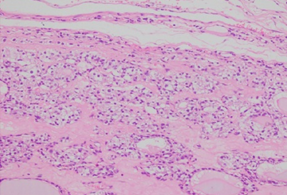
Introduction: Thyroid nodules represent a wide spectrum of neoplasms with different biological behaviors Majority of thyroid nodules are benign, but malignancy is found in approximately 5–15% of cases. Accurate diagnosis of these thyroid nodules is difficult, because of subtle and subjective histomorphological criteria. Immunohistochemistry method may play a complementary role to clarify diagnostic dilemma. CD56 is a neural cell adhesion molecule expressed on thyroid follicular cells. Down regulation of CD56 can shows correlation with tumour progression. Hector Battifora mesothelial epitope (HBME-1) is a membranous antigen located on follicular thyroid tumour cells and normal thyroid tissue is negative for HBME-1. Objective: To observe the expression of CD56 and HBME-1 in the diagnosis of surgically excised thyroid nodules. Methods: This cross sectional study was conducted in the Department of Pathology, Chittagong Medical College, Chattogram from March 2019 to February 2021. Immunohistochemistry was done at Armed Forces Institute of Pathology, Dhaka Cantonment. Sixty-three surgically resected thyroid nodules were evaluated to find out their histopathological type. Immunostaining was done by using primary antibody against CD56 (FLEX Monoclonal Mouse Anti-Human CD56 Clone 123C3 Ready to use (LINK). Denmark) and HBME-1 (Anti–Mesothelioma mouse monoclonal antibody HBME-1ab2383. Abcam, UK). Patient’s demographic data were collected and recorded in a predesigned data sheet. Statistical analysis was carried out as required. Ethical practice was ensured in every step of the study. Results: Among the 63 cases, mean age (±SD) of the patients was 39.47 ± 13.67 years and male to female ratio of 1:6.9. Thirty-four patients (76.3%) had multiple nodules. Among the 63 patients, 68.3% (43 cases) were histologically diagnosed as benign and 31 .7% (20 cases) as malignant thyroid nodules according 2017 WHO classification of thyroid tumours. Immunohistochemistry was performed using the markers CD56 and HBME-1 for all the 63cases.In present study weak to strong positive expression of CD56 was observed in 33(76.7%) cases out of 43 benign nodules whereas negative CD56 expression was observed in 20(100%) malignant cases. CD56 expression between benign and malignant lesion was statistically significant (p value, 0.002). HBME-1 was showed positive expression for 17(85%) out of 20 cases of malignant nodules and negative expression was observed in benign nodules. No statistically significant (p>0.250) difference was found between HBME-1 expression and histopathological diagnosis. So this study has improved the better understanding of thyroid nodules by expression of these immunomarkers (CD56 and HBME1) and thus may help the patients for selecting appropriate management protocol. Conclusion: In this study, Positive HBME-1 staining is a strong indicator of malignancy, although negative staining does not rule it out. IHC with CD56 and HBME-1 is considered to be important ancillary test in the diagnosis of thyroid neoplasms, but it does not replace the conventional histopathological examination.
Downloads
References
Saleh, H.A., Jin, B., Barnwell, J. and Alzohaili, O.,(2010). Utility of immunohistochemical markers in differentiating benign from malignant follicular- derived thyroid nodules. Diagnostic Pathology, 5(1).
Tamhane, S. and Gharib, H. (2016). Thyroid nodule update on diagnosis and management. Clinical Diabetes and Endocrinology, 2(1).
Hossain , M,A., Sarkar,M,J. , Dutta ,U,K., Karim,M,A. , Alam,Z,A.,(2014) Frequency of Malignancy in Solitary Thyroid Nodule and Multi-nodular Goitre. Bangladesh J Otorhinolaryngol 2014; 20(2): 55-65
Ozolins, A., Narbut, Z., Strumfa, I., Volanska, G., Stepanovs, K,. Gardovskis, J.,(2012) Immunohistochemical Expression of HBME-1, E- cadherinand CD56 in the Differential Diagnosis of Thyroid Nodules. , Medicina (Kaunas) 2012; 48(10):507-14.
Dirikoc, A., Faki, S., Baser, H., Özdemir, D., Aydin, C., Ersoy, R., Kilic, M., Kilicarslan, A. and Çakir, B. (2017). Thyroid malignancy risk in different clinical thyroid diseases. Turkish Journal of Medical Sciences, 47, 1509– 1519.
Topstad, D., Dickinson, J, A., (2017) Thyroid cancer incidence in Canada: a national cancer registry analysis. 10.9778/cmajo.20160162.
Naik, D., Jebasingh, Kf., and Thomas, N., (2018). Management of thyroid nodules in adults. Current Medical Issues, 16(2), 42.
Yao, Y., Chen, X., Wu, S., Liang, G., Zhang, H., Zhu, Q., et al., (2018). Thyroid nodules in centenarians: prevalence and relationship to lifestyle characteristics and dietary habits. Clinical Interventions in Aging, Volume 13, 515–522.
Vecchia,V,C,, Malvezzi ,M., Bosetti,C., Garavello,W. , Bertuccio,P., Levi ,F.,et al (2015).Thyroid cancer mortality and incidence: A global overview Int. J. Cancer: 136, 2187–2195.
Alshenawy, H., (2014). Utility of immunohistochemical markers in differential diagnosis of follicular cell-derived thyroid lesions. Journal of Microscopy and Ultrastructure, 2(3),127.
Muthusamy S. , Shah SH ., Suhaimi SNA ., Kassim N ., Mahasin M ., Saleh MFH., Isa NM (2018) CD56 expression in benign and malignant thyroid lesions Malaysia J Pathol40(2) . 111 – 119.
Schmidbauer, B., Menhart, K.,, Hellwig ,D., Grosse,J.,(2017) Differentiated Thyroid Cancer Treatment: State of the Art. Int.J.Mol.Sci.2017,18,1292.
Kakudo, K., K, Adel., Naggar, El., P, Steven., Hodak., Khanafshar, E., E, Yuri., Nikiforov., et al,.(2018) Noninvasive follicular thyroid neoplasm with papillary- like nuclear features (NIFTP) in thyroid tumor classification, 1–7 Available online at doi:10.1111/pin.12673.
Durmus, E.S., Ozcan, D., Yarikkaya, E., Kurt, A. and Arslan, A. (2016). CD56, HBME-1 and cytokeratin 19 expressions in papillary thyroid carcinoma and nodular thyroid lesions. Journal of Research in Medical Sciences, 21(1), 49.
Jiang, H., Tian, Y., Yan, W., Kong, Y., Wang, H., Wang, A., et al (2016). The Prevalence of Thyroid Nodules and an Analysis of Related Lifestyle Factors in BeijingCommunities.InternationalJournalofEnvironmentalResearchandPublic Health.
Yusuf, H., Rahman, A. K. M, Chowdhury F P., Mohiduzzaman, M., Banu, C.P., Sattar, M.A., et al(2008) Iodine deficiency disorders in Bangladesh, 2004-05: ten years of iodized salt intervention brings remarkable achievement in lowering goitre and iodine deficiency among children and women. Asia Pac J ClinNutr 2008;17 (4):620-628.
Jena, A., Patnayak, R., Prakash, J., Sachan, A., Suresh, V., and Lakshmi. A., (2015) Malignancy in solitary thyroid nodule: A clinicoradiopathological evaluation. 19(4): 498–503 Available online at doi:10.4103/2230-8210.159056.
Park, W.Y., Jeong, S. M., Lee ,JH., Kang ,HJ., Sin, DH., Choi ,KU., et al., (2009). Diagnostic value of decreased expression of CD56 protein in papillary carcinoma of the thyroid gland. Basic and Applied Pathology.2009; 2:63–8.
El, Demellawy .D., Nasr AL, Babay S,, Alowami, S,. (2009) Diagnostic utility of CD56 immunohistochemistry in papillary carcinoma of the thyroid. Pathol Res Pract.2009; 205(5):303–9.
Nasr, M.R, Mukhopadhyay, S., Zhang, S., Katzenstein, A.L., (2006). Immunohistochemical markers in diagnosis of papillary Thyroid carcinoma: Utility of HBME1 combined with CK19 immunostaining. Mod Pathol. 2006; 19 (12):1631–7.